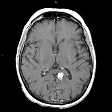
Magnetic resonance imaging (MRI) uses radiofrequency waves and a strong magnetic field rather than x-rays to provide remarkably clear and detailed pictures of internal organs and tissues.
The patient is placed on a sliding table and positioned comfortably for the MRI examination, which will generally take 15-45 minutes, depending on how many images are needed. Patients are asked to remain still during the actual imaging process (only a few seconds to a few minutes at a time), but between sequences some movement is allowed.
The technique has proven very valuable for the diagnosis of a broad range of pathologic conditions in all parts of the body including cancer, heart and vascular disease, stroke, and joint and musculoskeletal disorders. MRI requires specialized equipment and expertise and allows evaluation of some body structures that may not be as visible with other imaging methods.
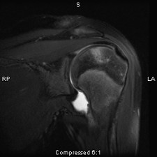
Because MRI can give such clear pictures of soft-tissue structures near and around bones, it is the most sensitive exam for spinal and joint problems. MRI is widely used to diagnose sports-related injuries, especially those affecting the foot, ankle, knee, shoulder, hip, elbow and wrist. The images allow the physician to see even very small tears and injuries to ligaments and muscles.
In addition, MRI of the heart, aorta, coronary arteries and blood vessels is a fast, noninvasive tool for diagnosing coronary artery disease and heart problems. Physicians can examine the size and thickness of the chambers of the heart and determine the extent of damage caused by a heart attack or progressive heart disease.
Organs of the chest and abdomen—including the lungs, liver, kidney, spleen, pancreas and abdominal vessels—can also be examined in high detail with MRI, enabling the diagnosis and evaluation of tumors and functional disorders. MRI is growing in popularity as an alternative to traditional x-ray mammography in the early diagnosis of breast cancer. Because no radiation exposure is involved, MRI is often the preferred diagnostic tool for examination of the male and female reproductive systems, pelvis and hips and the bladder. Breast MRI is also used in certain cases as an adjunctive study to Mammography.