Advances in the field of breast imaging have saved many lives. Cancers that were not detected in the past can be found at earlier stages, resulting in improved survival.
The triad of self-breast exam, physical exam and mammography still are the best ways to detect breast cancer early. New techniques of diagnosis and biopsy are continually evolving, enabling women to make decisions early in the fight against breast cancer.
Mammography
Mammography remains the basic test to detect the earliest, pre-invasive, form of breast cancer. Annual screening mammography is generally recommended for all women 40 years and over. Full field digital mammography is the same test as the older film screen form of mammography except in how the radiographic information is collected and displayed. Electronic manipulation of digital images has been shown to improve the accuracy of mammography in younger women and those with dense breasts.
Mammography is the only screening tool for breast cancer that is known to reduce deaths due to breast cancer through early detection. Even so, mammograms do not detect all breast cancers. Some breast lesions and abnormalities are not visible or are difficult to interpret on mammograms. Medical studies are currently being done to determine whether other imaging methods can help supplement mammography by detecting small breast cancers that may not be visible with mammography.
Breast Ultrasound
Breast ultrasound, also called sonography, has become increasingly beneficial in the work-up of breast abnormalities. Using the hand held transducer, like a rectangular microphone, an ultrasound exam can be directed right to a breast abnormality that has been detected on self- or physical exam. Frequently, an abnormality on mammography will be further evaluated with sonography. Ultrasound is useful in determining if the abnormality is a cyst or a solid structure as well as other characteristics. Specific criteria are used to judge the potential for malignancy, and if suspicious, ultrasound may be used to direct a biopsy.
Ultrasound exams are usually completed in about 30 minutes. You will be asked to lie on a padded table with the arm of the side of interest resting overhead. Sometimes, you may be asked to sit up or roll to one side or the other with cushion behind your back for support. Warmed gel on the skin is used as an interface between the hand-held scanning devise. The ultrasonographer will obtain multiple images for review by the radiologist. The excess gel is wiped off after the exam.
Breast MRI
Magnetic resonance imaging (MRI) is a non-invasive way of viewing organs, soft tissues, bone and other internal body structures without the use of x-rays. MRI uses a magnetic field and radio waves together with a computer to create cross-sectional, three-dimensional pictures of the head and body. MRI of the breast is not a replacement for mammography or ultrasound imaging but rather is a supplemental tool for detecting and staging breast cancer and other breast abnormalities.
Medical studies are currently being conducted to determine whether MRI and other imaging methods can contribute to the early detection of, and prevention of deaths from, breast cancer. The American Cancer Society is now recommending annual breast MRI in addition to annual mammography for women at high risk, defined as 20-25% or greater lifetime risk of breast cancer.
Because an MRI exam of the breast requires intravenous contrast material, a technologist will insert an intravenous (IV) line into a vein in your hand or arm. You will be positioned face down on the moveable table with your breasts hanging into the cushioned openings and your arms overhead. The table will then be moved into the magnet of the MRI unit. The technologist will leave the room while the MRI examination is performed. You will be asked to lie still while the machine acquires the images. Imaging is done in sequences, each lasting between one and fifteen minutes. You will know when images are being recorded because you will hear tapping or thumping sounds when the coils that create the magnetic field are turned on. After an initial series of scans, the contrast material is injected into the intravenous line. Additional series of images are taken following the injection. Holding still is very important in making the images match up. When your exam is completed, you will wait a short time on the table while the images are evaluated to ensure no additional images are needed. Your intravenous line will then be removed. The imaging session lasts about hour and the total exam will take approximately an hour and a half. Processing of the data for review by the radiologist will take additional time, after you have left.
Image Guided Biopsy
Radiologic imaging has made great strides in the early detection of abnormalities that may represent breast cancer but imaging alone cannot diagnose a cancer. For that, a biopsy must be performed for interpretation by a pathologist. When a lump is felt in the breast, biopsy can be done directly on that lump. For breast abnormalities detected only on radiologic images, that imaging will be needed to direct the site of biopsy. Sampling of tissue exactly at the site of concern is paramount to accurate diagnosis. Once a biopsy is done, it usually takes a couple days for pathology to complete the processing and interpretation of the specimen. There are times when confident sampling is not accomplished and a surgical biopsy may be necessary.
Many women ask if these techniques actually spread the tumor. There is no evidence that this happens, and women should be comfortable knowing that a diagnosis can be made in a minimally-invasive fashion that provides her with accurate information needed to deal with options available to treat breast cancer.
Stereotactic Mammotome Biopsy
Mammographically guided biopsy is typically done with stereotaxis. This requires a specialized table with a mammography x-ray tube that swings 15 degrees either side of center and is interfaced with a computer. The target for biopsy is identified on each image by the radiologist and the computer calculates the location in 3 dimensions.
You will be asked to lie face down on the table with the breast for biopsy hanging through the opening in the table and placed in compression. Images will be obtained for the radiologist to identify the area of concern and calculate the coordinates of biopsy. Your breast will then be cleansed and the site of puncture numbed with local anesthetic. Additional images will be acquired during the placement of the needle to assess positioning for biopsy. The biopsy is done using a vacuum assisted automated devise operated by the radiologist. Multiple samples are obtained to give the pathologist enough to make a sound judgment. More images may be done to further assess adequacy of the biopsy. If calcifications are being sampled, the specimen will also be x-rayed. Often, a marker is placed to indicate the site of biopsy. The marker is tiny, made of MRI-safe metal and has a distinctive appearance on future mammograms. Once an adequate biopsy has been determined, the devise will be withdrawn and pressure held at the puncture site followed by placement of a bandage. A mammogram is usually done at the completion of the procedure. You will be given instruction on care of the puncture. The procedure usually takes about an hour to an hour and a half.
Ultrasound Guided Biopsy
Ultrasound guidance for biopsy will often be requested for suspicious masses detected on mammography and/or well demonstrated on the sonogram. Because the procedure is quicker and done with the patient in a more comfortable position, ultrasound guidance for biopsy is preferred if technically possible.
You will be asked to lie on the same table that the initial ultrasound exam was performed on and in a similar position as during that first exam. The ultrasound exam will be repeated using the same gel to re-demonstrate the abnormality and determine an approach to biopsy. The biopsy approach is usually at a distance from the site of the abnormality to allow good visualization of the lesion by the transducer while it is sampled. The skin at the site of approach is then cleansed and numbed with local anesthetic. Additional anesthetic is administered along the expected track of the biopsy needle. Often an introducing device will be placed to the margin of the abnormality allowing multiple passes with the biopsy needle. The ultrasound transducer in a sterile cover is simultaneously held over the site of the abnormality while the biopsy is being performed to insure correct placement. The biopsy devise may be a spring loaded needle or vacuum assisted; both acquire cores of tissue. The goal is to obtain an adequate sample for the pathologist to interpret. Once adequate sampling has been determined, pressure will be held at the puncture site and a bandage applied. The procedure usually takes about an hour.
MRI Guided Biopsy
MRI requires a strong magnetic field, limiting the kinds of instruments that can be brought into the scanning room. The face down position required for MRI of the breasts is probably the least comfortable and creates an awkward position for possible biopsy. For these reasons, efforts will be made to seek adequate visualization of an MRI detected abnormality by ultrasound or mammography to allow a more facile biopsy. However, there are some cancers that can only be detected on MRI and capability for MRI guided biopsy should be in place whenever a scan is performed.
You will be asked to lie face down on the same MRI table, but this time there will be paddles gently compressing the breast to be biopsied. The MRI detected abnormality may only be demonstrated with intravenous injection of the contrast material and, if so, this will need to be repeated in preparation for the biopsy. Series of images will be acquired, similar to your initial exam, to allow for identification of the abnormality and calculation of the coordinates for biopsy. The skin over the site to be biopsied will be cleansed and numbed with local anesthetic. An introducing device will be placed and more images acquired to determine correct site of biopsy. Multiple core samples will then be taken, using either a spring-loaded needle or a vacuum assisted devise. Additional images may be needed to determine the adequacy of sampling. A marker may be placed at the biopsy site. When the procedure is finished, pressure is held at the puncture site and a dressing applied. A mammogram may be performed after the procedure. The procedure will take about one to two hours.
Aftercare
You will be given instructions on care of you breast following a biopsy by whatever means. An ice pack for the first day helps control pain and swelling. A good supportive bra or sports bra will help to limit strain at the biopsy site. A Tylenol type medication should be all that is needed for pain. The area will be bruised and tender but will gradually improve over a couple weeks.
Image Guided Localization
For biopsy of an unknown or to guide lumpectomy for treatment of a cancer, your surgeon may request radiologic localization. This may be done using mammography or ultrasound.
Mammographic localization is done using mammography with a grid as the compression paddle. The site of interest is identified on the mammogram and a needle is place to that site through the grid. Additional mammograms are done to confirm that the needle is in the right place. Ultrasound guidance is similar, placing the needle to the site of concern and confirming positioning using ultrasonography. Depending on the surgeon’s preference, a small amount of blue dye or a thin wire, or both, are then placed and the needle is removed. If there is a wire, the excess outside the skin will be taped down. You will then go to lymphoscintigraphy or to have your procedure done.
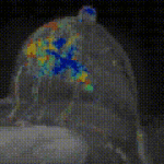
Lymphoscintigraphy
Sentinel node biopsy may be performed at the time of your lumpectomy. A small amount of radioactive material will need to be administered in a manner that guides your surgeon to the first lymph nodes draining the lymphatic channels of the breast. Placing this material in the superficial layers of the skin optimizes uptake by the lymphatics. The radioactive material is mixed with a local anesthetic and injected, usually at the areolar margin of the breast. This is usually done in the Nuclear Medicine department so images confirming lymphatic and lymph node uptake can be acquired using specialized radioactivity-detecting cameras. This procedure will follow the localization procedure, if you have one done. After lymphoscintigraphy, you will go to surgery.
For more detailed information on this examination, visit RadiologyInfo.